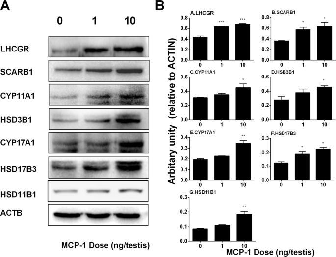
Morphology of Leydig cells in the testes after in vivo MCP-1 treatment.
Por um escritor misterioso
Last updated 04 abril 2025

IJMS, Free Full-Text
Monocyte Chemoattractant Protein-1 stimulates the differentiation of rat stem and progenitor Leydig cells during regeneration, BMC Developmental Biology
Frontiers Insights into the Development of the Adult Leydig Cell Lineage from Stem Leydig Cells

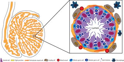
The Sertoli cell: one hundred fifty years of beauty and plasticity - França - 2016 - Andrology - Wiley Online Library

Morphology of Leydig cells in the testes after in vivo MCP-1 treatment.
Cells, Free Full-Text

Morphology of Leydig cells in the testes after in vivo PTHrP
Frontiers Sertoli Cell Immune Regulation: A Double-Edged Sword

Effect of TNF on testosterone production. Leydig cells were cultured in

Human testicular peritubular cells: more than meets the eye in: Reproduction Volume 145 Issue 5 (2013)
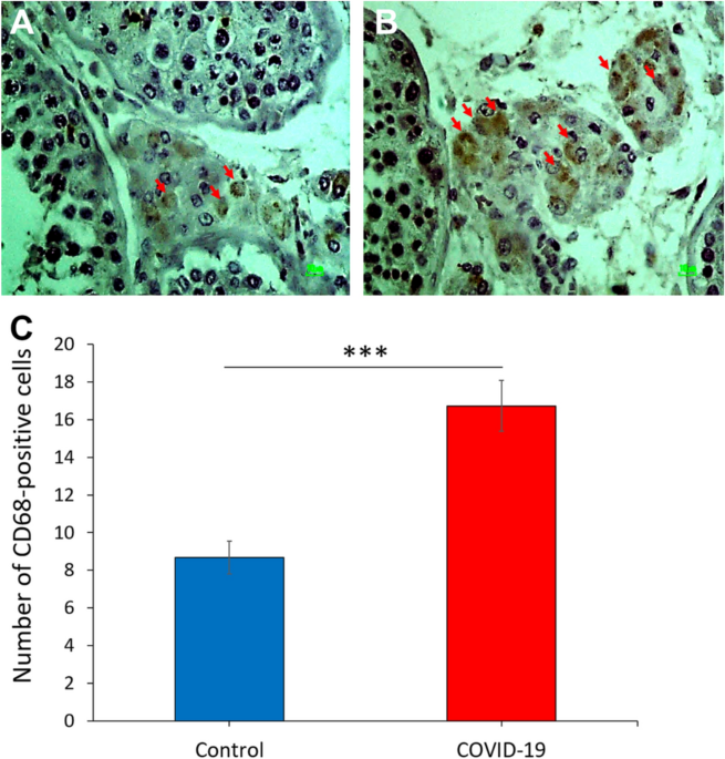
COVID-19 disrupts the blood–testis barrier through the induction of inflammatory cytokines and disruption of junctional proteins
Monocyte Chemoattractant Protein-1 stimulates the differentiation of rat stem and progenitor Leydig cells during regeneration, BMC Developmental Biology
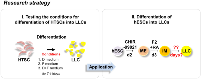
Rapid Differentiation of Human Embryonic Stem Cells into Testosterone-Producing Leydig Cell-Like Cells In vitro

Morphology of Leydig cells in the testes after in vivo PTHrP
Recomendado para você
-
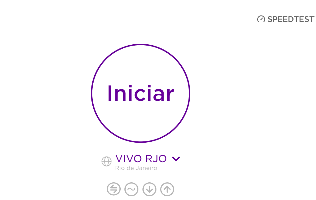 Teste de Velocidade Vivo, Teste Vivo, Power, Internet04 abril 2025
 -
 Teste de Velocidade Vivo, Contrate Online04 abril 2025
 -
 Teste Anpad 2023 - Aula 03 de matemática financeira do curso ao vivo04 abril 2025
 -
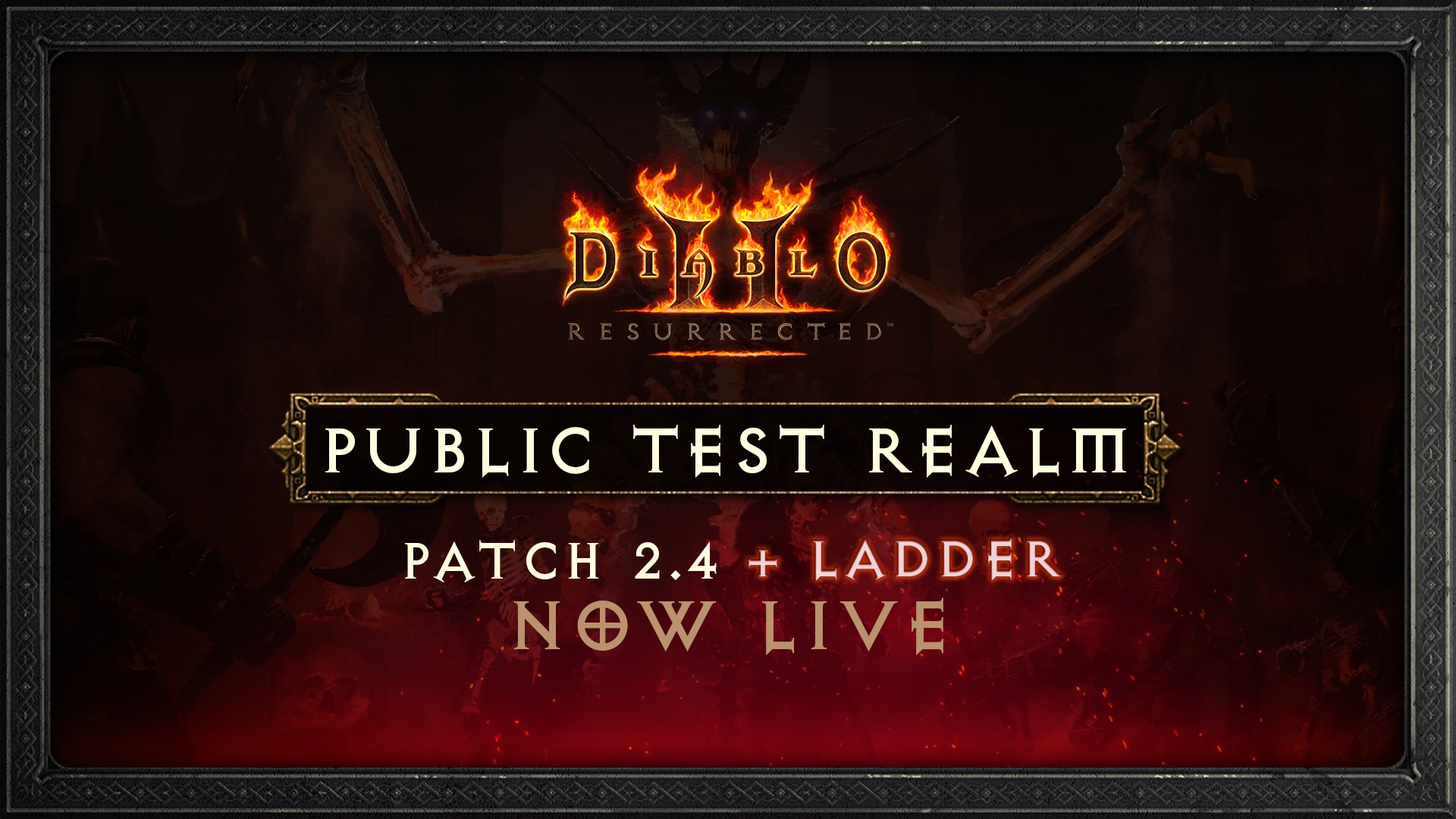 RTP do Patch 2.4 de Diablo II: Resurrected, Teste Competitivo04 abril 2025
 -
 Sobre Um Fundo Vermelho Vivo Um Cartão De Cor De Madeira Leve Com Um Teste De Massa Ilustração Stock - Ilustração de palavra, mensagem: 23630853204 abril 2025
 -
 Vivo X100 ganha imagens oficiais, amostras de fotos e testes de desempenho - Canaltech04 abril 2025
 -
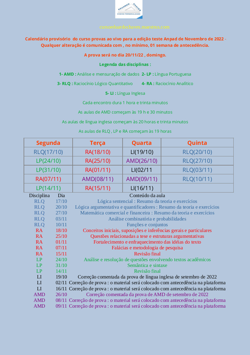 Teste Anpad 2022 - 2023 - Curso de provas ao vivo para a edição de novembro de 202204 abril 2025
 -
LinkedIn lança teste ao vivo de postagens de IA generativa04 abril 2025
-
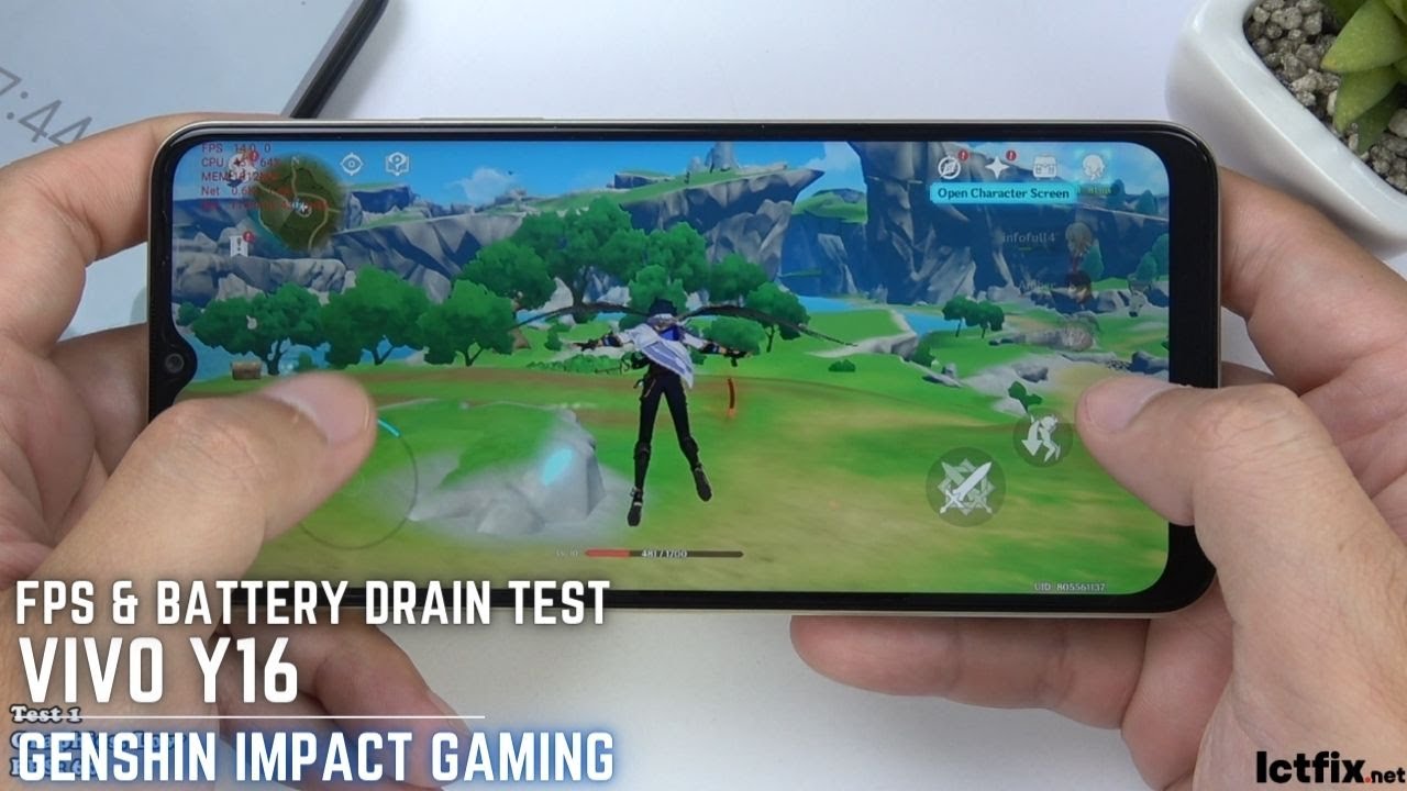 Vivo Y16 Genshin Impact Gaming test04 abril 2025
 -
 Teste Padrão Vivo Da Cauda Do Crocodilo Do Corpo Vivo Para O Fundo Foto de Stock - Imagem de bens, cultivar: 10243900204 abril 2025

você pode gostar
-
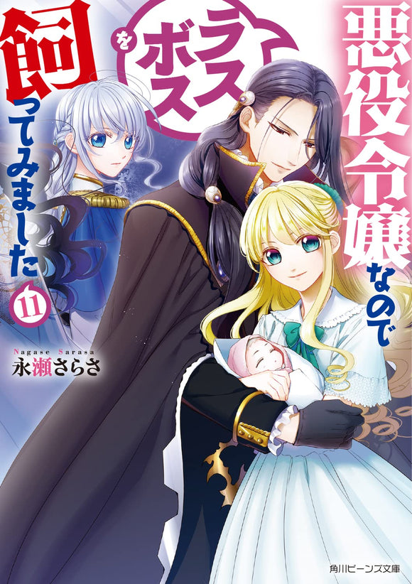 I'm the Villainess, So I'm Taming the Final Boss (Akuyaku Reijou nanode Last Boss wo Kattemimashita) 11 (LIght Novel) – Japanese Book Store04 abril 2025
 -
format(webp)) ENTREVISTA: Luísa Viotti, a voz brasileira de Makima em Chainsaw04 abril 2025
ENTREVISTA: Luísa Viotti, a voz brasileira de Makima em Chainsaw04 abril 2025 -
 I Ranked All Yu-Gi-Oh! 5D's Characters In a Tier List! - YGO Tier List Video04 abril 2025
 -
 How to fix controller not working in God of War PC04 abril 2025
 -
 Travis Kelce Reacts to Having Taylor Swift at Chiefs vs. Jets Game04 abril 2025
 -
 SAIA CARGO - Ventury Store - Nara Martins04 abril 2025
 -
 Download do APK de Pegar personagens do Roblox para Android04 abril 2025
 -
 Chess Quote: Anatoly Karpov Chess quotes, Anatoly karpov, History quotes04 abril 2025
 -
 Evil West (Multi-Language) for PlayStation 404 abril 2025
 -
 Stream Maestro Luiz Fernando da Costa Listen to Dobrados Em festa - Sociedade Cultural e Musical de Santo Amaro playlist online for free on SoundCloud04 abril 2025