Figure 1. [The normal human retina fundus]. - Webvision - NCBI
Por um escritor misterioso
Last updated 01 abril 2025
![Figure 1. [The normal human retina fundus]. - Webvision - NCBI](https://www.ncbi.nlm.nih.gov/books/NBK554706/bin/Archetecture_Fovea-Image006.jpg)
The normal human retina fundus photo shows the optic nerve (right), blood vessels and the position of the fovea (center).
![Figure 1. [The normal human retina fundus]. - Webvision - NCBI](https://ars.els-cdn.com/content/image/1-s2.0-S266646902300026X-gr2.jpg)
Cell death mechanisms in retinal phototoxicity - ScienceDirect
![Figure 1. [The normal human retina fundus]. - Webvision - NCBI](https://www.mdpi.com/cells/cells-12-01987/article_deploy/html/images/cells-12-01987-g001.png)
Cells, Free Full-Text
![Figure 1. [The normal human retina fundus]. - Webvision - NCBI](https://media.springernature.com/lw685/springer-static/image/art%3A10.1186%2Fs13024-023-00655-y/MediaObjects/13024_2023_655_Fig1_HTML.png)
Retinal ganglion cell repopulation for vision restoration in optic neuropathy: a roadmap from the RReSTORe Consortium, Molecular Neurodegeneration
![Figure 1. [The normal human retina fundus]. - Webvision - NCBI](https://www.ncbi.nlm.nih.gov/books/NBK1222/bin/retinoschisis-Image001.jpg)
Figure 1. [Fundus photo of a male]. - GeneReviews® - NCBI Bookshelf
![Figure 1. [The normal human retina fundus]. - Webvision - NCBI](http://webvision.instead-technologies.com/wp-content/uploads/2014/07/armdretina-300x270.jpeg)
11.2 The Electroretinogram and Electrooculogram: Clinical Applications. by Donnell Creel – Webvision
![Figure 1. [The normal human retina fundus]. - Webvision - NCBI](https://www.ncbi.nlm.nih.gov/books/NBK11556/bin/factsf2a.gif)
Facts and Figures Concerning the Human Retina - Webvision - NCBI Bookshelf
![Figure 1. [The normal human retina fundus]. - Webvision - NCBI](https://www.cell.com/cms/attachment/2119048143/2088495043/gr1.jpg)
Cell-Based Therapy for Degenerative Retinal Disease: Trends in Molecular Medicine
![Figure 1. [The normal human retina fundus]. - Webvision - NCBI](http://webvision.med.utah.edu/imageswv/glaucretina.jpeg)
Simple Anatomy of the Retina : 네이버 블로그
![Figure 1. [The normal human retina fundus]. - Webvision - NCBI](https://www.ncbi.nlm.nih.gov/books/NBK590568/bin/oca-oa-ov-Image001.jpg)
Figure 1. [(A) Illustration of the unique]. - GeneReviews® - NCBI Bookshelf
![Figure 1. [The normal human retina fundus]. - Webvision - NCBI](https://www.frontiersin.org/files/Articles/1106728/fpubh-11-1106728-HTML/image_m/fpubh-11-1106728-g001.jpg)
Frontiers Biomechanical homeostasis in ocular diseases: A mini-review
![Figure 1. [The normal human retina fundus]. - Webvision - NCBI](http://webvision.org.es/wp-content/uploads/2017/01/Fig01.png)
Retinal Degeneration, Remodeling and Plasticity. Bryan William Jones, Robert E. Marc and Rebecca L. Pfeiffer - Webvision
Recomendado para você
-
 Retina - Definition and Detailed Illustration01 abril 2025
 -
 Retina Raleigh, Diabetic Retinopathy Raleigh01 abril 2025
 -
 Descolamento de Retina - Instituto de Moléstias Oculares01 abril 2025
 -
 Specialty Eye Care What is a Retina Specialist? - Specialty Eye Care01 abril 2025
 -
 Descolamento de retina: sintomas e tratamento01 abril 2025
 -
 Retina Conditions Tallahassee, What is the Retina?01 abril 2025
 -
 Retina International01 abril 2025
 -
 Structure and Function of the Eyes - Eye Disorders - Merck Manuals01 abril 2025
 -
 Descolamento de retina pode afetar até 3% das crianças – Jornal da USP01 abril 2025
 -
 Cirurgia e Tratamento para Descolamento de Retina - COHR01 abril 2025
você pode gostar
-
 Gaming Website Template - Free PSD - Freebie Supply01 abril 2025
 -
 A Spider-Man 2 Secret Room Might Be Hinting At Daredevil DLC - GameSpot01 abril 2025
 -
 Reitoria IFRJ, 2014-2016: Reitoria faz um panorama da gestão, Edição nº 0101 abril 2025
 -
 Yagate Kimi ni Naru / Opening HD Legendado PT/BR01 abril 2025
 -
 Goku ssj6 HD wallpapers01 abril 2025
 -
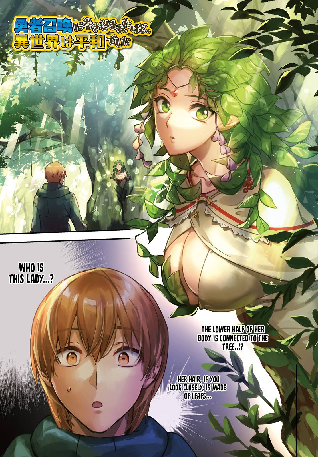 Read Yuusha Shoukan ni Makikomareta kedo, Isekai wa Heiwa deshita01 abril 2025
 -
 IJM Land launches brand new business hub in Bandar Rimbayu01 abril 2025
 -
 garfo no espanhol - dicionário Português-Espanhol01 abril 2025
 -
 Gukesh dethrones Vishy Anand's 37-year-long reign as India no.1 officially, also becomes World no.8 - ChessBase India01 abril 2025
 -
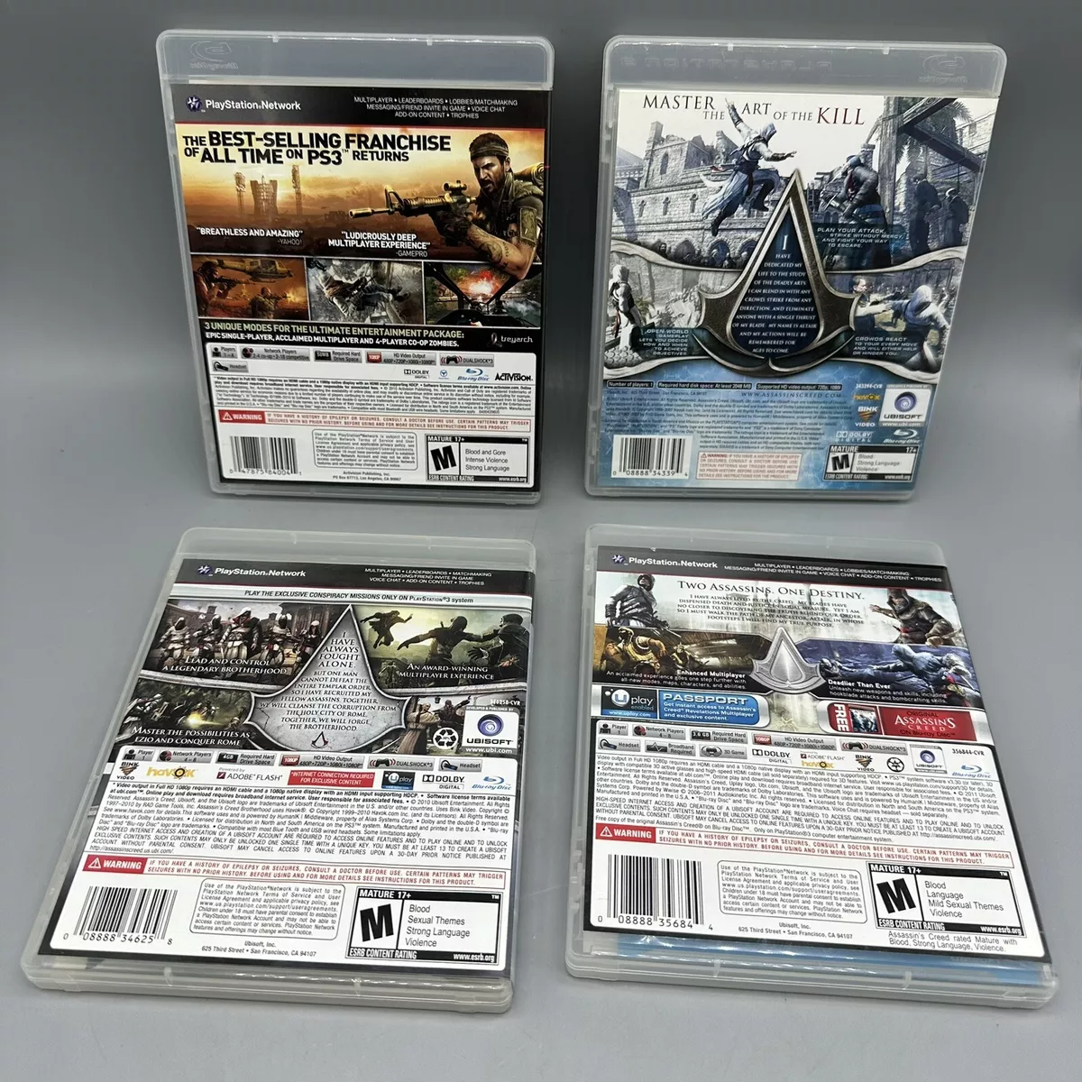 Lot of 4 Ps3 Games - Assassins Creed, Brotherhood, Revelations, Call Of Duty Bl01 abril 2025