Figure 1 from Brain surface temperature under a craniotomy.
Por um escritor misterioso
Last updated 05 abril 2025

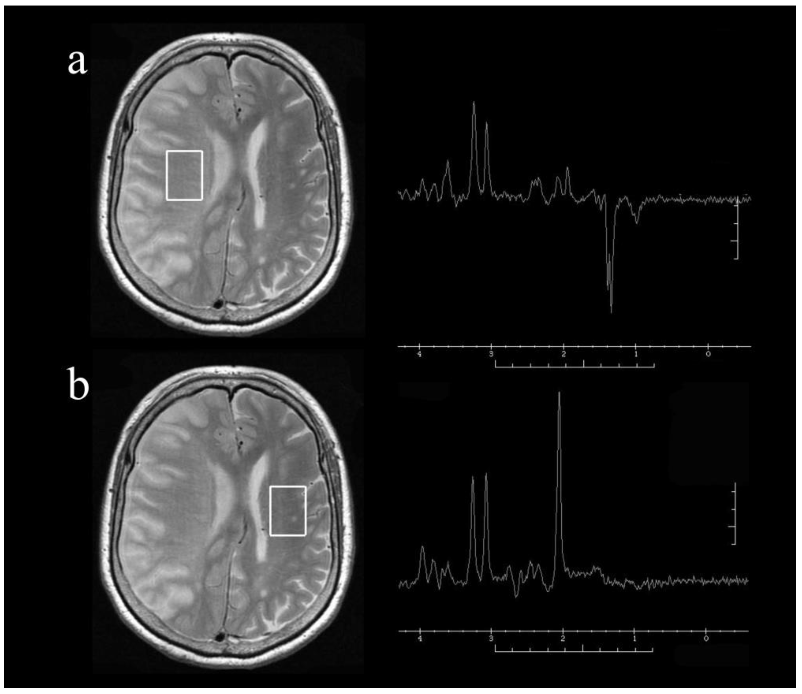
Fig. 1. Rapid cooling of the brain surface in an in vivo mouse preparation. A: schematic representation of a cranial window during recording of temperature and single-cell activity in the anesthetized mouse. The main potential routes of heat transfer are indicated. B: brain surface temperature measured with the thermocouple during replacement of the artificial cerebrospinal fluid (ACSF) with fresh ACSF warmed to 38°C. ACSF was replaced twice, indicated by the arrowheads. - "Brain surface temperature under a craniotomy."
Recording of pig neuronal activity in the comparative context of the awake human brain
Sensors, Free Full-Text
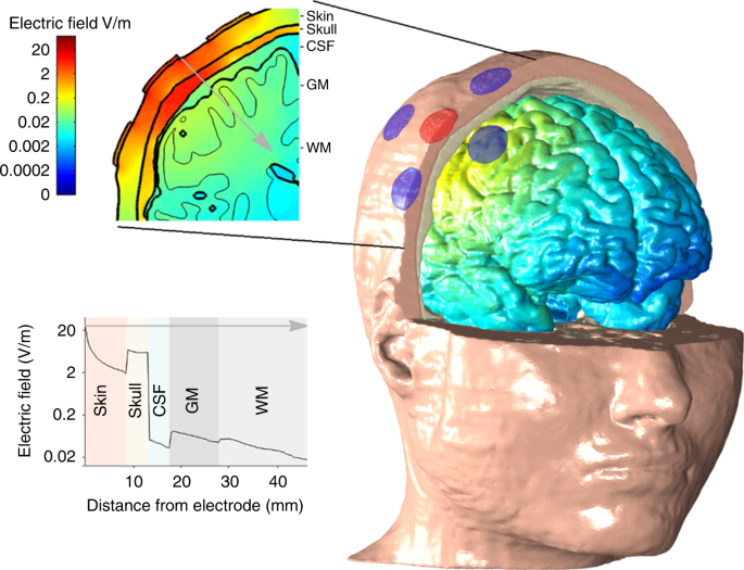
tACS motor system effects can be caused by transcutaneous stimulation of peripheral nerves
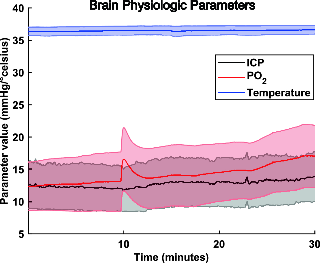
Regional pressure and temperature variations across the injured human brain: comparisons between paired intraparenchymal and ventricular measurements, Critical Care

JFB, Free Full-Text

Therapeutic Hypothermia And Neuroprotection

Intraoperative detection of blood vessels with an imaging needle during neurosurgery in humans
Figure 1 from Brain surface temperature under a craniotomy.

Craniotomy, Expert Surgeon

Infrared thermography display of cortical temperature in cats
Recomendado para você
-
 Brain Test Level 367 It's cold the fireplace needs more fire in 202305 abril 2025
 -
 Tech Thursday: Voice-to-Text - by Breana Bayraktar05 abril 2025
 -
 Association of body mass index and waist-to-hip ratio with brain structure05 abril 2025
 -
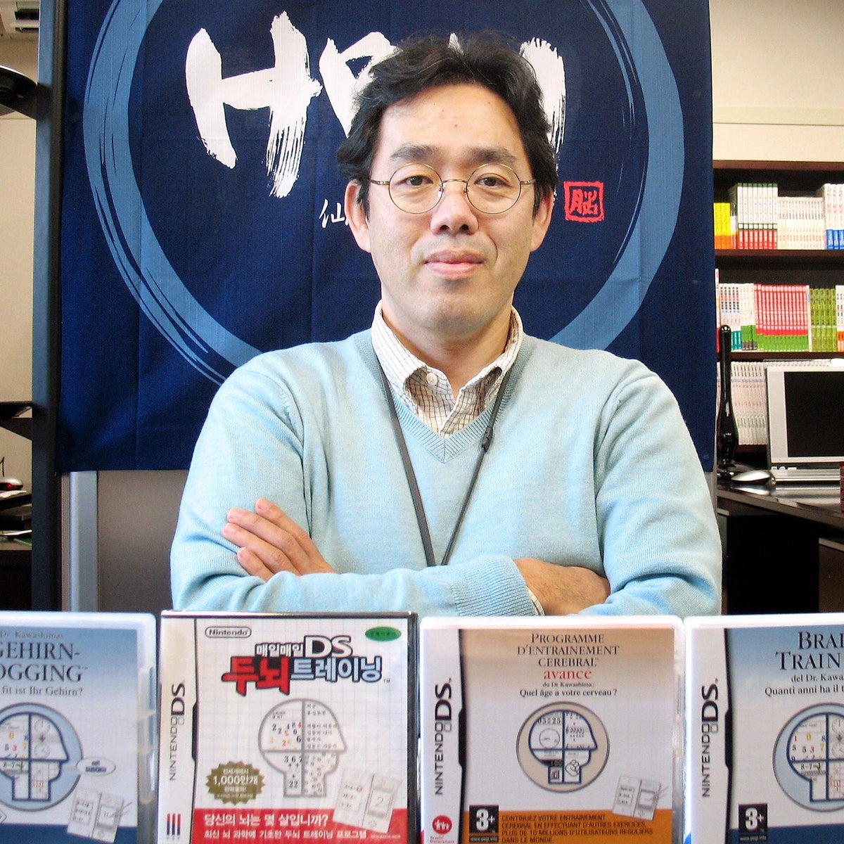 Ryuta Kawashima: The devil who cracked the dementia code, The Independent05 abril 2025
 -
 Saatnya mencari cuan! (Brain Test Level 367) - CadeMedia05 abril 2025
 -
 Neuroimaging and deep learning for brain stroke detection - A review of recent advancements and future prospects - ScienceDirect05 abril 2025
 -
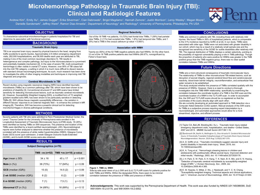 PDF) Microhemorrhage Pathology in Traumatic Brain Injury (TBI): Clinical and Radiologic Features05 abril 2025
 -
A Cell Type Selective YM155 Prodrug Targets Receptor-Interacting Protein Kinase 2 to Induce Brain Cancer Cell Death05 abril 2025
-
 Effectiveness of management strategies for uninvestigated dyspepsia: systematic review and network meta-analysis05 abril 2025
 -
 TMS Reveals Dynamic Interaction between Inferior Frontal Gyrus and Posterior Middle Temporal Gyrus in Gesture-Speech Semantic Integration05 abril 2025

você pode gostar
-
 Leagues Cup 2023: Teams, groups, schedule, Lionel Messi debut05 abril 2025
 -
The Humble Monthly Stream featuring Assassin's Creed Origins05 abril 2025
-
 Nanatsu no Taizai revela sequência 'Four Knights of the Apocalypse', adaptação do mangá; confira o trailer05 abril 2025
 -
 Jogue com 20 Números na Lotofacil Acumulada em 5.000.000,0005 abril 2025
 -
 UFC 274: Michael Chandler delivers devastating knockout of Tony05 abril 2025
 -
bichos lore05 abril 2025
-
USPGameDev São Paulo SP05 abril 2025
-
 D4C: Love Train, Your Bizarre Adventure Wiki05 abril 2025
 -
 Sudoku.game 🕹️ Play on CrazyGames05 abril 2025
 -
 Pokemon Articuno, Pokemon Galarian, Articuno Plush, Doll05 abril 2025