Assessment of Myocardial Viability Using Nuclear Medicine Imaging in Dextrocardia
Por um escritor misterioso
Last updated 03 abril 2025
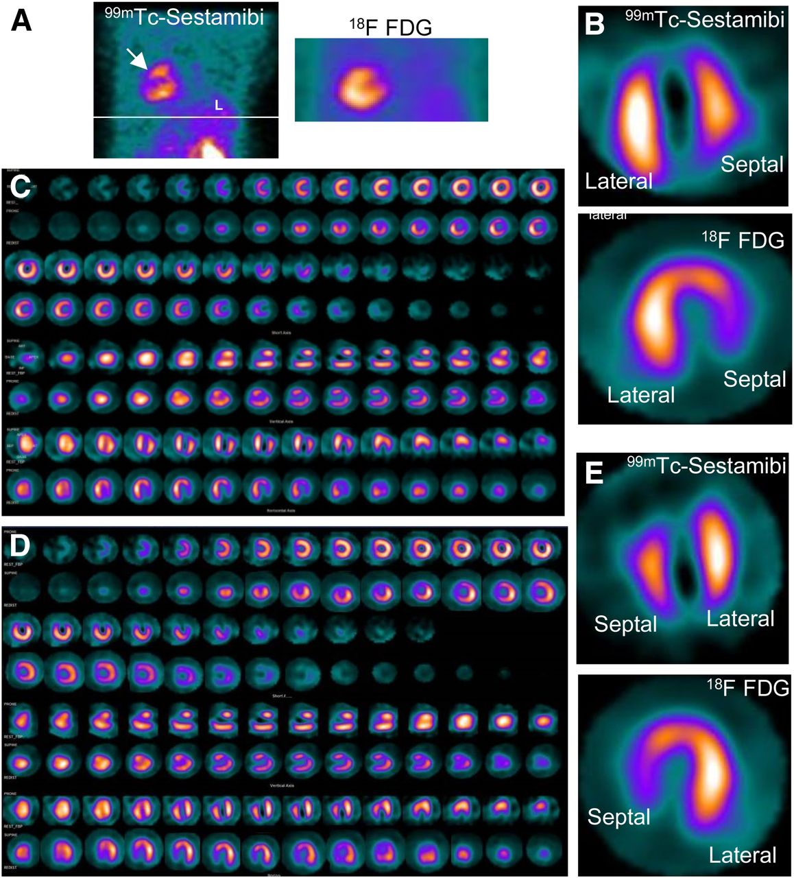
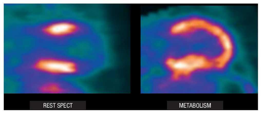
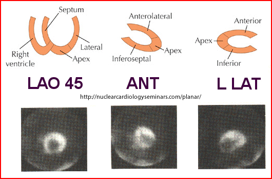
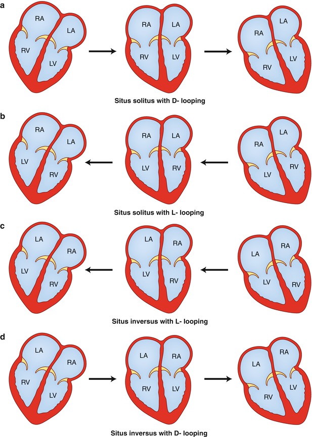
Imaging of dextrocardia in humans requires an understanding of the orientation of the heart chambers and walls. There are many types of cardiac malpositioning, such as dextrocardia (with or without situs inversus), mesocardia, and levocardia. Myocardial perfusion scintigraphy of dextrocardia has been explained in case reports and imaging atlases; however, myocardial viability assessment using nuclear medicine imaging techniques is less documented in the literature. Methods: In 2 cases of dextrocardia with situs inversus and 1 case of mesocardia, myocardial viability was assessed using 99mTc-sestamibi rest perfusion scintigraphy and 18F-FDG PET. Cardiac SPECT images of dextrocardia with situs inversus were acquired using the feet-first supine position with a 180° arc from left anterior oblique to right posterior oblique, whereas a right-lateral–to–left-lateral arc was used for mesocardia. The processing and reconstruction were done by entering the dataset for the feet-first supine position and repeating after entering the dataset for the feet-first prone position. The 2 sets of reconstructed images were compared for orientation of walls and cardiac chambers. Results: The first processing, using the feet-first supine position, revealed an interchanged septum and lateral wall in reconstructed images of dextrocardia with situs inversus. This interchange was corrected by changing the position to prone during processing of the rest perfusion and PET raw data. The display of cardiac slices in various axes matched the conventional nomenclature for the septum and lateral wall, leading to easy interpretation. However, this change was not required in the mesocardia, for which the location of the heart chambers was not interchanged. Conclusion: Because the acquisition protocol for SPECT is a semicircular orbit, the various types of dextrocardia require careful selection of the arc, with the patient positioning kept feet-first supine. Processing and reconstruction of data by changing the patient position to prone was found to be most useful method of matching the septum and lateral wall orientation for interpretation of images.
Assessment of Myocardial Viability Using Nuclear Medicine Imaging in Dextrocardia

SPECT myocardial perfusion imaging in patients with Dextrocardia

Society for Cardiovascular Magnetic Resonance/European Society of Cardiovascular Imaging/American Society of Echocardiography/Society for Pediatric Radiology/North American Society for Cardiovascular Imaging Guidelines for the Use of Cardiac Magnetic

Approach to Dextrocardia in Adults: Review
Ischemic Cardiomyopathy: A Clinical Nuclear Cardiology Perspective
Artifacts and Pitfalls in Myocardial Perfusion Imaging
Levocardia disease: Malacards - Research Articles, Drugs, Genes, Clinical Trials

State of the Art: Imaging for Myocardial Viability: A Scientific Statement From the American Heart Association
Nuclear Cardiology acquisition Protocols
Visceroatrial Situs in Congenital Heart Disease
Perfusion Measurements of the Myocardium: Radionuclide Methods and Related Techniques
Pharmaceuticals, Free Full-Text
Recomendado para você
-
 Brain Test Level 372 Walkthrough03 abril 2025
 -
 Он хочет быть выше. 372 уровень Brain Test03 abril 2025
 -
 Brain Test Level 202 Solve the puzzle in 202303 abril 2025
 -
 Western blots show p65 antibodies that passed the test of specificity03 abril 2025
 -
 Brain Test Level 371 372 373 374 375 Walkthrough03 abril 2025
 -
The Hop Shoppe03 abril 2025
-
 Number Tic-Tac-Toe IQ Puzzle on the App Store03 abril 2025
 -
 Class Notes for PY 372 at University of Alabama (UA)03 abril 2025
 -
Brain Sciences Center - News03 abril 2025
-
MTVBaseAfrica03 abril 2025


você pode gostar
-
 Bebe Reborn Pode Tomar Banho Tem O Cabelo Fio A Fio Realista03 abril 2025
 -
 Lost Life - Act 1: Broken - Gameplay03 abril 2025
 -
is it takes two crossplay|TikTok Search03 abril 2025
-
 Memórias e Imagens: Cabra-cega03 abril 2025
 -
 Motocicleta De Desenho Animado Bonito Clássico, Vista Lateral, Isolado. Royalty Free SVG, Cliparts, Vetores, e Ilustrações Stock. Image 15134065203 abril 2025
 -
 Moana' Live Action Cast: Will 'The Rock' Play as Maui? Latin Post - Latin news, immigration, politics, culture03 abril 2025
 -
 The Lords (German band) - Wikipedia03 abril 2025
 -
Domestic na Kanojo Chapter 40 Discussion - Forums03 abril 2025
-
 Jogos e brincadeiras em apps para crianças de 2 e 3 anos // Renata Conrado03 abril 2025
 -
 Crunchyroll on X: Good Morning ~ (via Golden Time) / X03 abril 2025