Studying Virus Replication with Fluorescence Microscopy
Por um escritor misterioso
Last updated 12 abril 2025
The results from research on SARS-CoV-2 virus replication kinetics, adaption capabilities, and cytopathology in Vero E6 cells, done with the help of fluorescence microscopy, are described in this article.
Studying Virus Replication with Fluorescence Microscopy, Science Lab

Single-Virus Tracking: From Imaging Methodologies to Virological Applications
Microscopy Deep Learning Predicts Viral Infections - Automatic detection of virus-infected cells - solely on the fluorescence of the cell nucleus
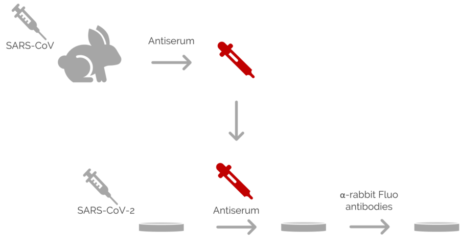
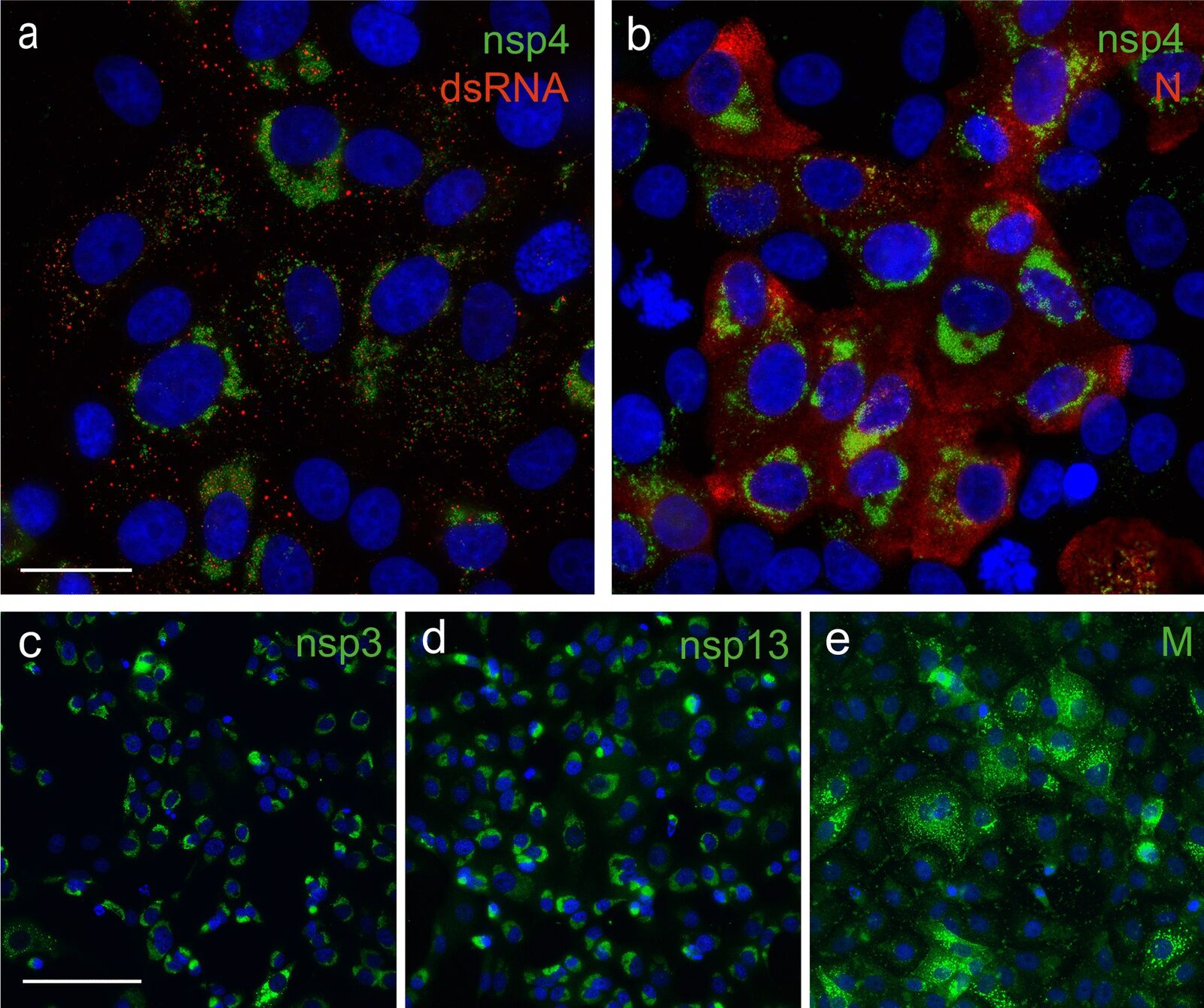
Frontiers Imaging Flow Cytometry and Confocal Immunofluorescence Microscopy of Virus-Host Cell Interactions

Virus morphology: Insights from super-resolution fluorescence microscopy - ScienceDirect
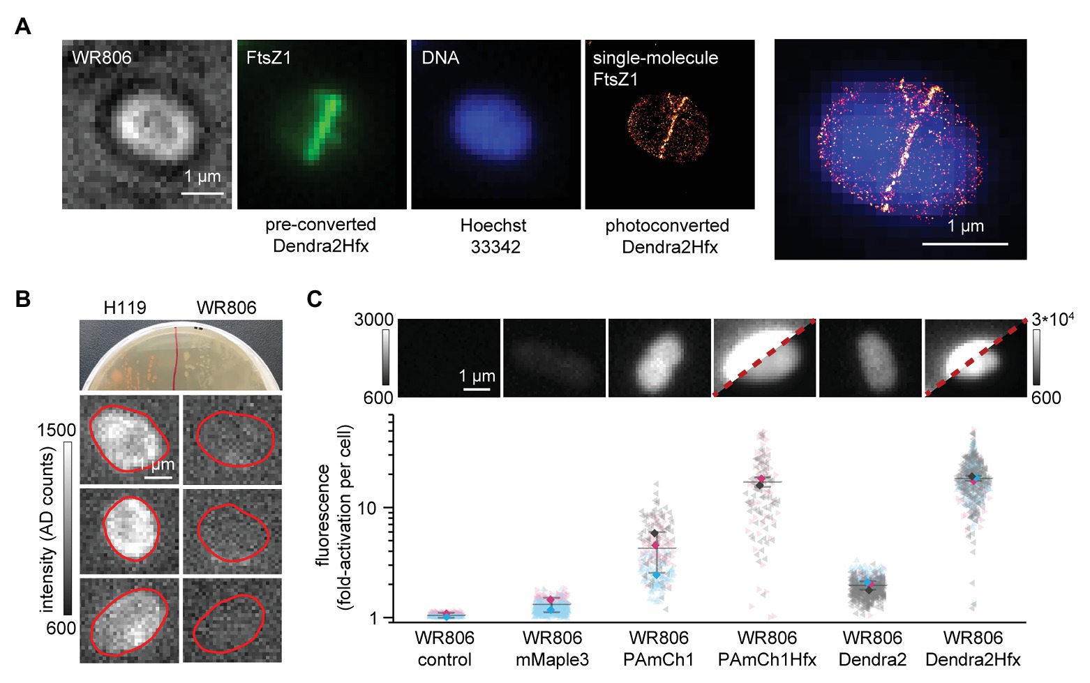
Frontiers Establishing Live-Cell Single-Molecule Localization Microscopy Imaging and Single-Particle Tracking in the Archaeon Haloferax volcanii

Microscopy deep learning predicts virus infections and reveals mechanics of lytic-infected cells - ScienceDirect

Viral replication visualized under fluorescence microscopy
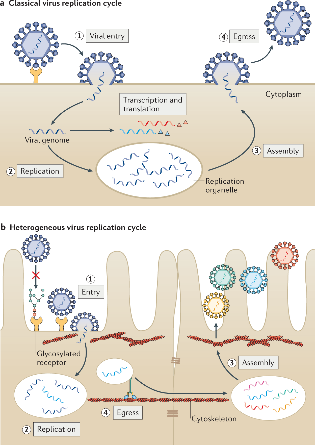
Viral and host heterogeneity and their effects on the viral life cycle
Recomendado para você
-
 Top 74 Similar websites like steamunlocked.pro and alternatives12 abril 2025
 -
 People Playground Download PC Game Full Version12 abril 2025
 -
 helping (ahumanbeing8246) on how to download people playground12 abril 2025
 -
 Does SteamUnlocked have Trojans : r/SteamUnlocked12 abril 2025
 -
People Playground download link in comments #peopleplayground12 abril 2025
-
 Purplis Sandbox Free Download (v0.1.7) » STEAMUNLOCKED12 abril 2025
 -
 Free Running Free Download in 202312 abril 2025
 -
 People FIGHT in a Underground Facility - People Playground12 abril 2025
 -
 How to apply Steam Workshop items to cracked People Playground v12 abril 2025
 -
 Enjoy Great Online Sports With the Mobile App in 202312 abril 2025

você pode gostar
-
 Shigeru Miyamoto: site reúne acervo com mais de 450 entrevistas do criador de Mario - Game Arena12 abril 2025
 -
 Dinossauro Tiranossauro Rex - Schleich12 abril 2025
 -
 Chashu Pork (Marinated Braised Pork Belly) - Closet Cooking12 abril 2025
 -
![[30 Tales From the Foundation] SCP New Series (Tales from the Foundation New Collection)](https://m.media-amazon.com/images/W/MEDIAX_792452-T2/images/I/61IbcNu1+6L._AC_UF1000,1000_QL80_.jpg) [30 Tales From the Foundation] SCP New Series (Tales from the Foundation New Collection)12 abril 2025
[30 Tales From the Foundation] SCP New Series (Tales from the Foundation New Collection)12 abril 2025 -
 Jogo de quebra-cabeça para crianças - cidade dos desenhos animados12 abril 2025
 -
 Cómo reunir las Bolas de Dragon y qué deseo pedir en Dragon Ball Xenoverse 212 abril 2025
 -
 Spring Fens Kenai Watershed Forum12 abril 2025
 -
 Cemu v1.8.012 abril 2025
 -
 Roblox Crashando e saindo do Jogo Como Resolver12 abril 2025
 -
 ALL NEW *SECRET* CODES in ANIME WARRIORS CODES! (Roblox Anime12 abril 2025